Human Skeletal System Drawing at Explore collection of Human Skeletal
Human Anatomy - Skeleton. Click on the labels below to find out more about your skeleton. More human anatomy diagrams: front view of muscles, back view of muscles, organs, nervous system. Assemble.
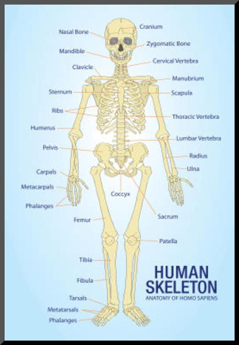
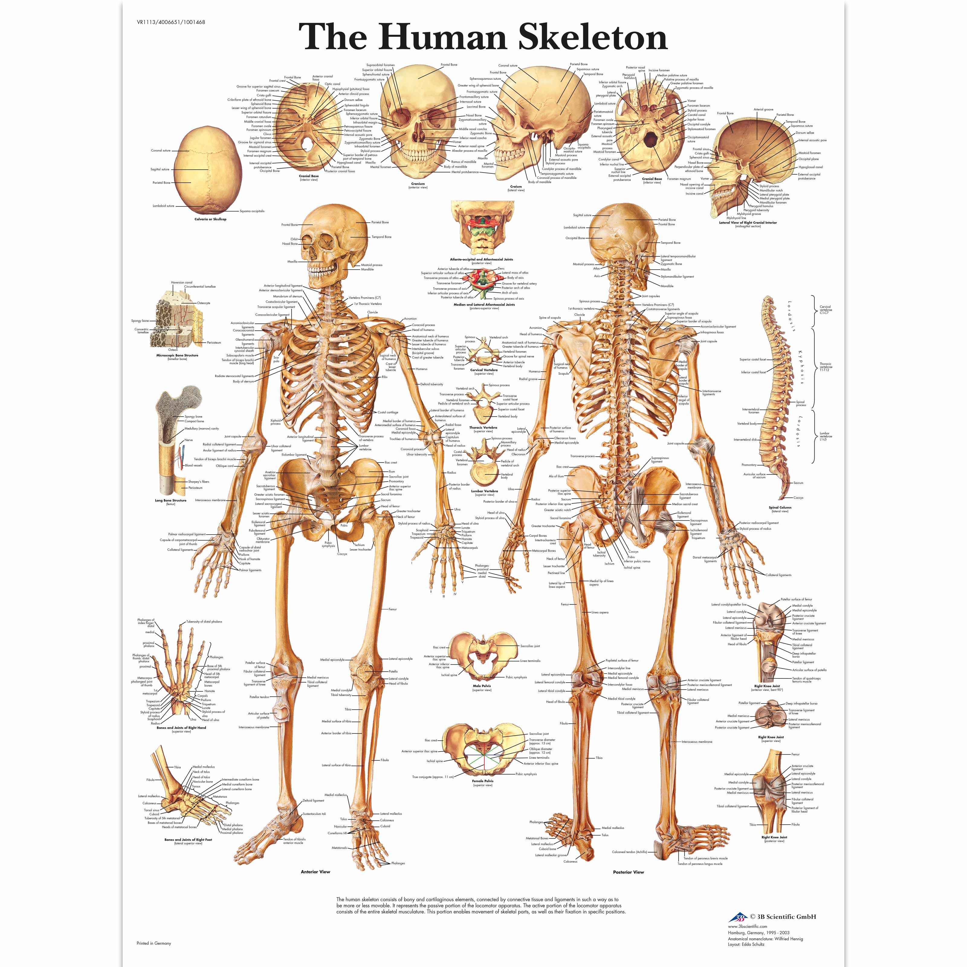
Human Skeleton Anatomy Anatomical Chart Poster Print Mounted Print 13x19
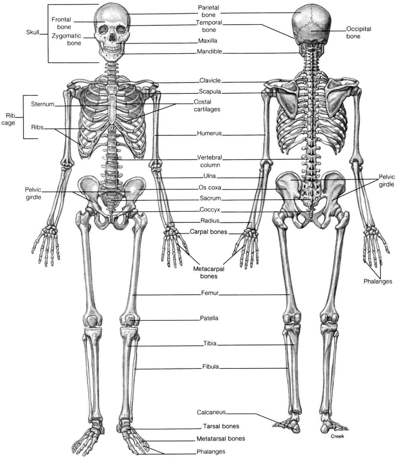
In adults, the skeletal system includes 206 bones, many of which are shown in Figure 14.2.2 14.2. 2. Bones are organs made of dense connective tissues, mainly the tough protein collagen. Bones contain blood vessels, nerves, and other tissues. Bones are hard and rigid due to deposits of calcium and other mineral salts within their living tissues.
Human Skeletal System Drawing at Explore collection of Human Skeletal
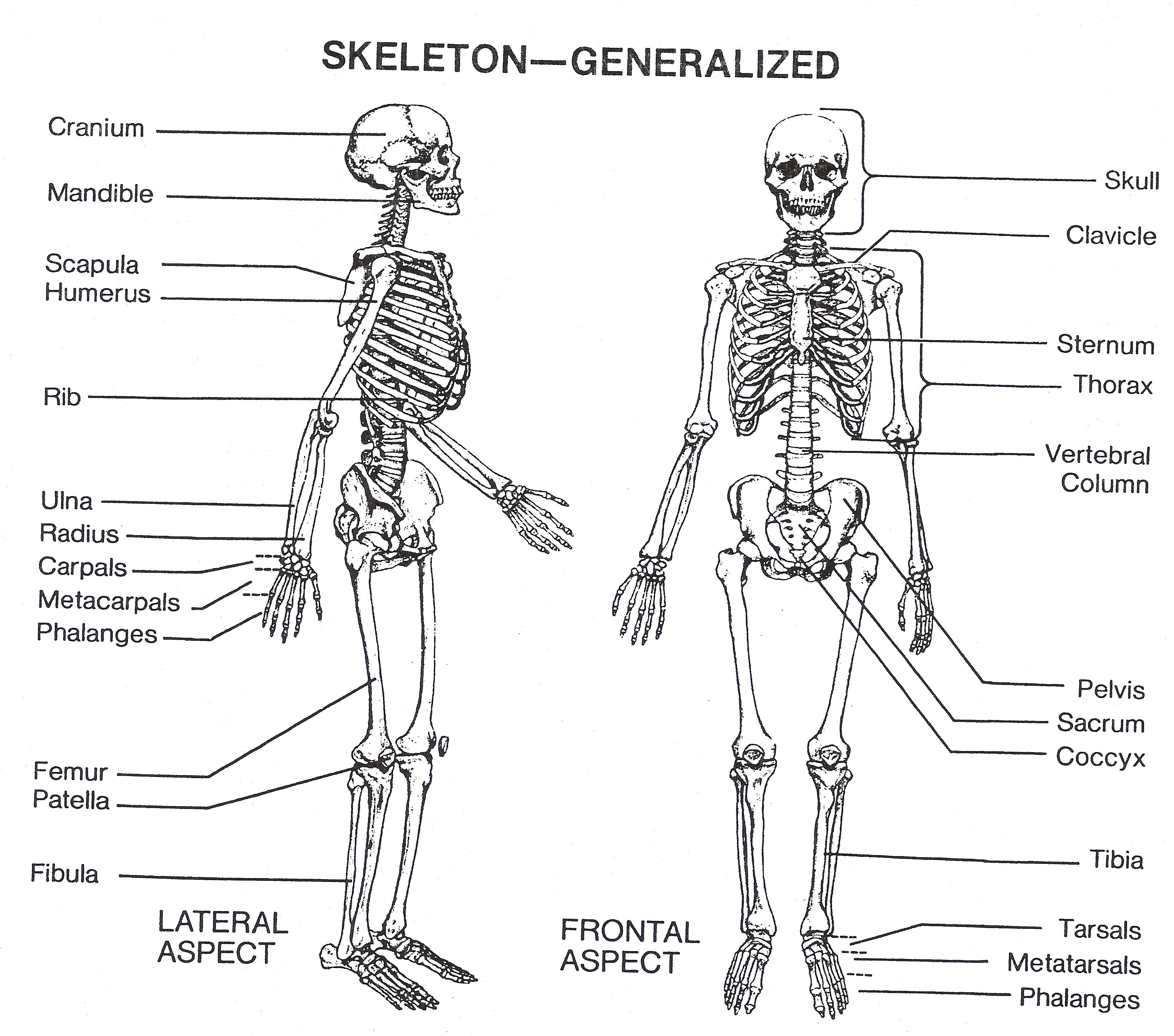
A diagram of the human skeleton showing bone and cartilage. Protection of the heart, lungs, and other organs and structures in the chest creates a problem somewhat different from that of the central nervous system. These organs, the function of which involves motion, expansion, and contraction, must have a flexible and elastic protective covering.
Images 04. Skeletal System Basic Human Anatomy
Skeletal System The skeletal system includes all of the bones and joints in the body. Muscular System The muscular system is responsible for the movement of the human body. Cardiovascular System The cardiovascular system consists of the heart, blood vessels, and the approximately 5 liters of blood that the blood vessels transport.
Human Skeleton Poster Human Skeleton Chart Paper
The human skeleton is the internal framework of the human body. It is composed of around 270 bones at birth - this total decreases to around 206 bones by adulthood after some bones get fused together. The bone mass in the skeleton makes up about 14% of the total body weight (ca. 10-11 kg for an average person) and reaches maximum mass between the ages of 25 and 30.
Human Skeletal System Diagram Health Images Reference
3. The Skeleton Protects Vital Organs. The brain is surrounded by bones that form part of the skull. The heart and lungs are located within the thoracic cavity, and the vertebral column provides structure and protection for the spinal cord. 4. Interactions Between the Skeleton, Muscles, and Nerves Move the Body.

7 Structure of the skeleton. Image reproduced with permission from... Download Scientific Diagram
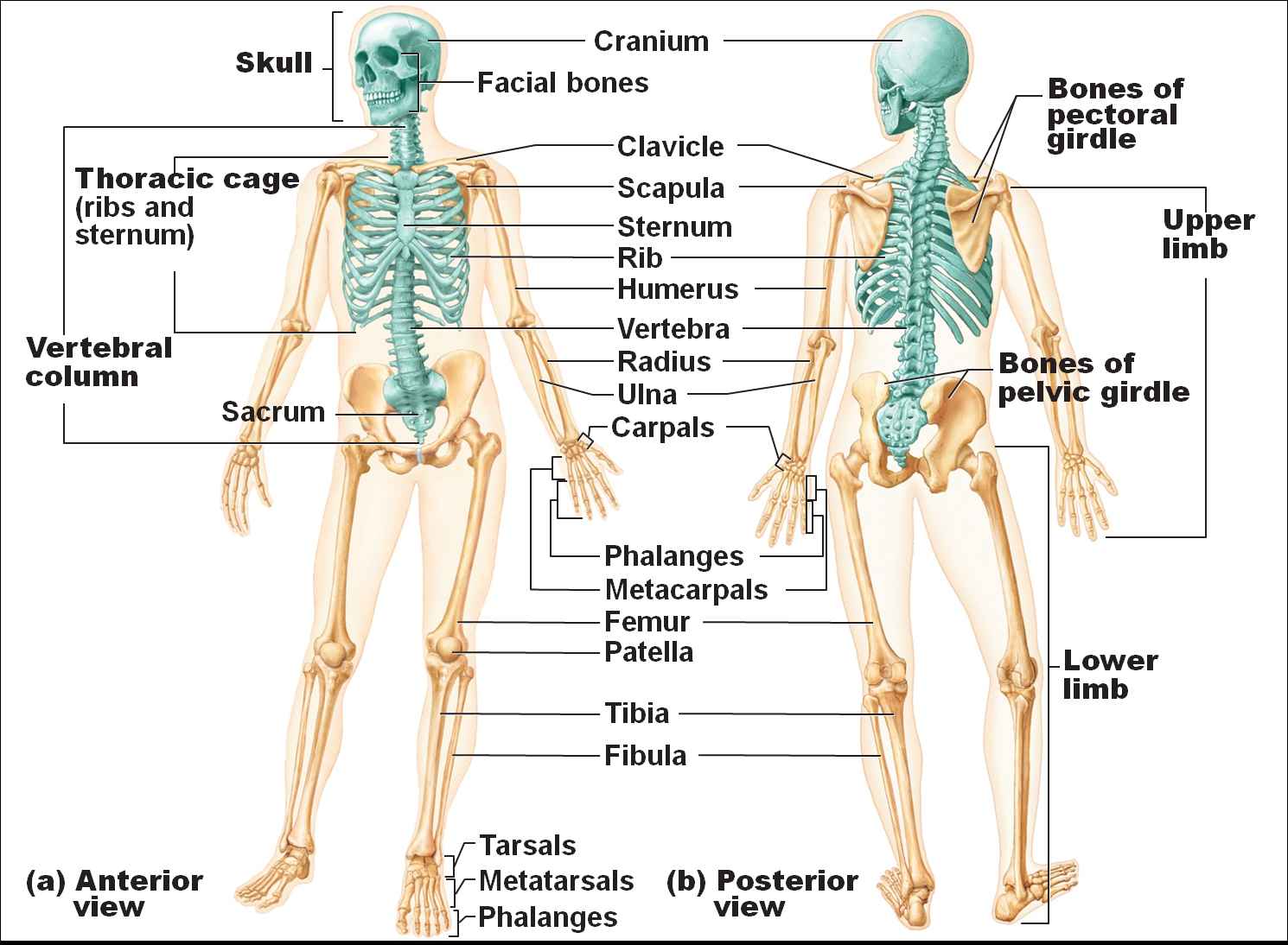
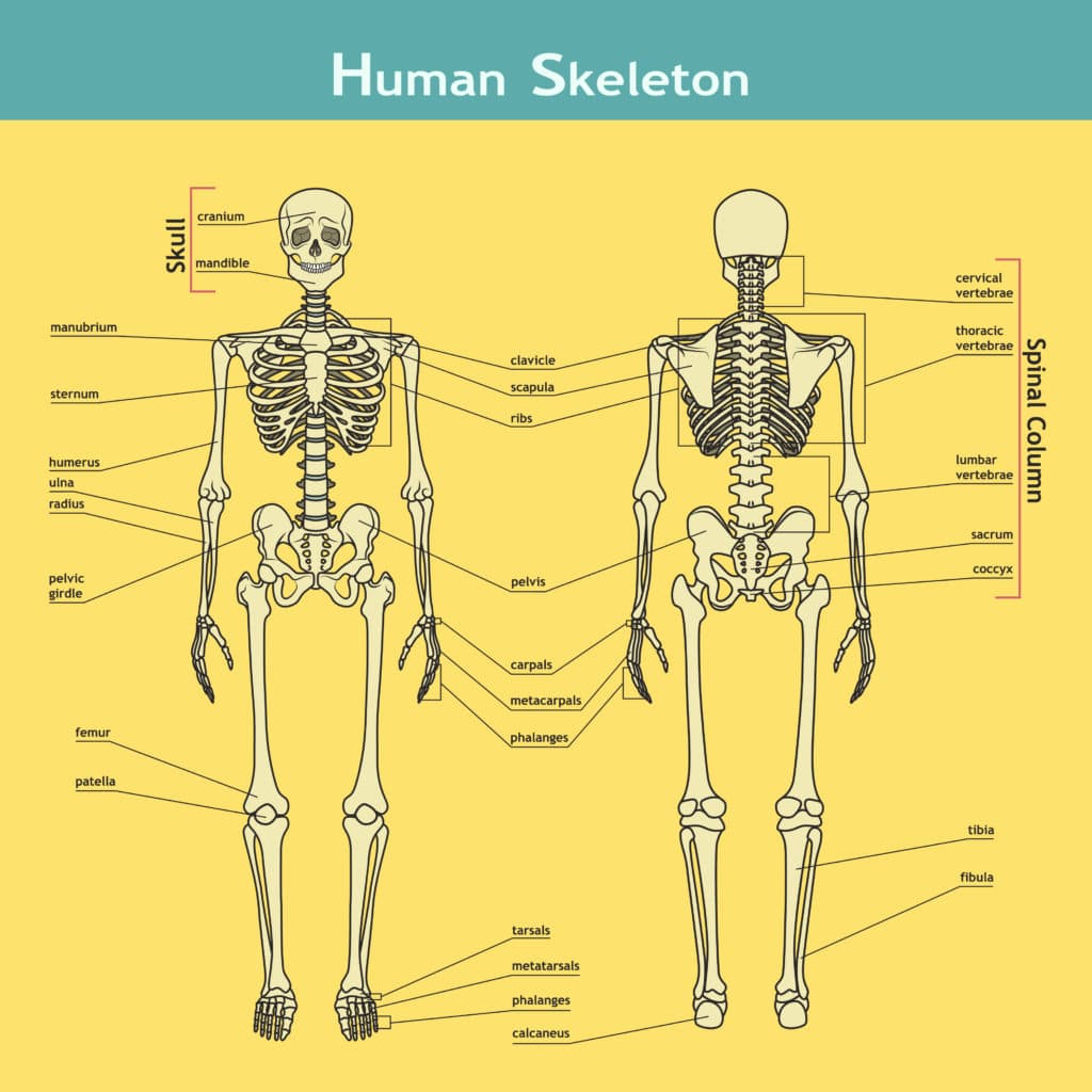
Here's a skeletal system diagram providing you with a broad overview of the two skeletons and the bones in the body: [Main bones of the skeletal system (anterior view)] The axial skeleton is essentially the midline, or central core region, and consists of the bones of the skull (cranium) together with the bones of the trunk.
skeletal system diagram
The skeletal system is your body's central framework. It consists of bones and connective tissue, including cartilage, tendons, and ligaments. It's also called the musculoskeletal system. Advertisement. Cleveland Clinic is a non-profit academic medical center. Advertising on our site helps support our mission.
Skeleton diagram
skeleton, the supportive framework of an animal body.The skeleton of invertebrates, which may be either external or internal, is composed of a variety of hard nonbony substances.The more complex skeletal system of vertebrates is internal and is composed of several different types of tissues that are known collectively as connective tissues.This designation includes bone and the various fibrous.

The Skeletal System Diagram Labeled koibana.info Human anatomy, Human skeleton, Human
The human skeletal system consists of all of the bones, cartilage, tendons, and ligaments in the body. Altogether, the skeleton makes up about 20 percent of a person's body weight. An adult's.

Skeleton Anatomy Poster Skeletal System Anatomical Chart Company
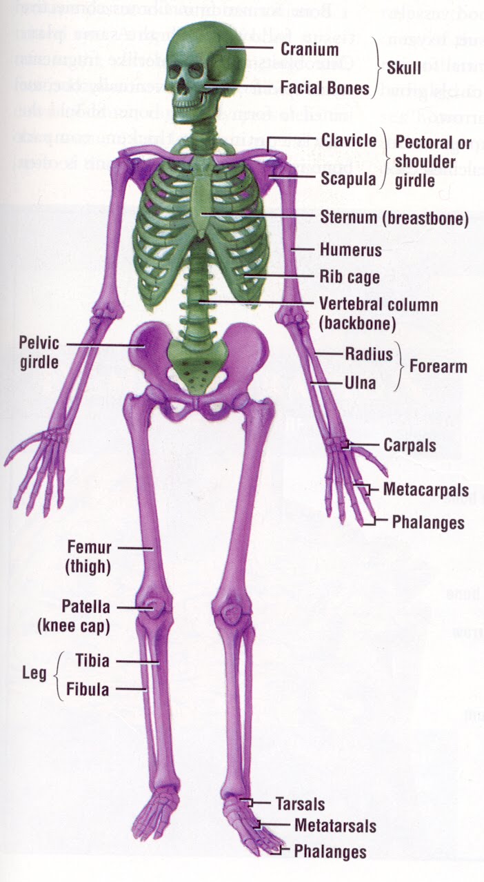
The skeletal system is made up of your bones, ligaments, and cartilage. Though its main function is to provide structural support for the body, it also stores important minerals—such as calcium—forms red blood cells, and protects your internal organs. The skeletal system can break down into two main categories—the axial skeleton, which.
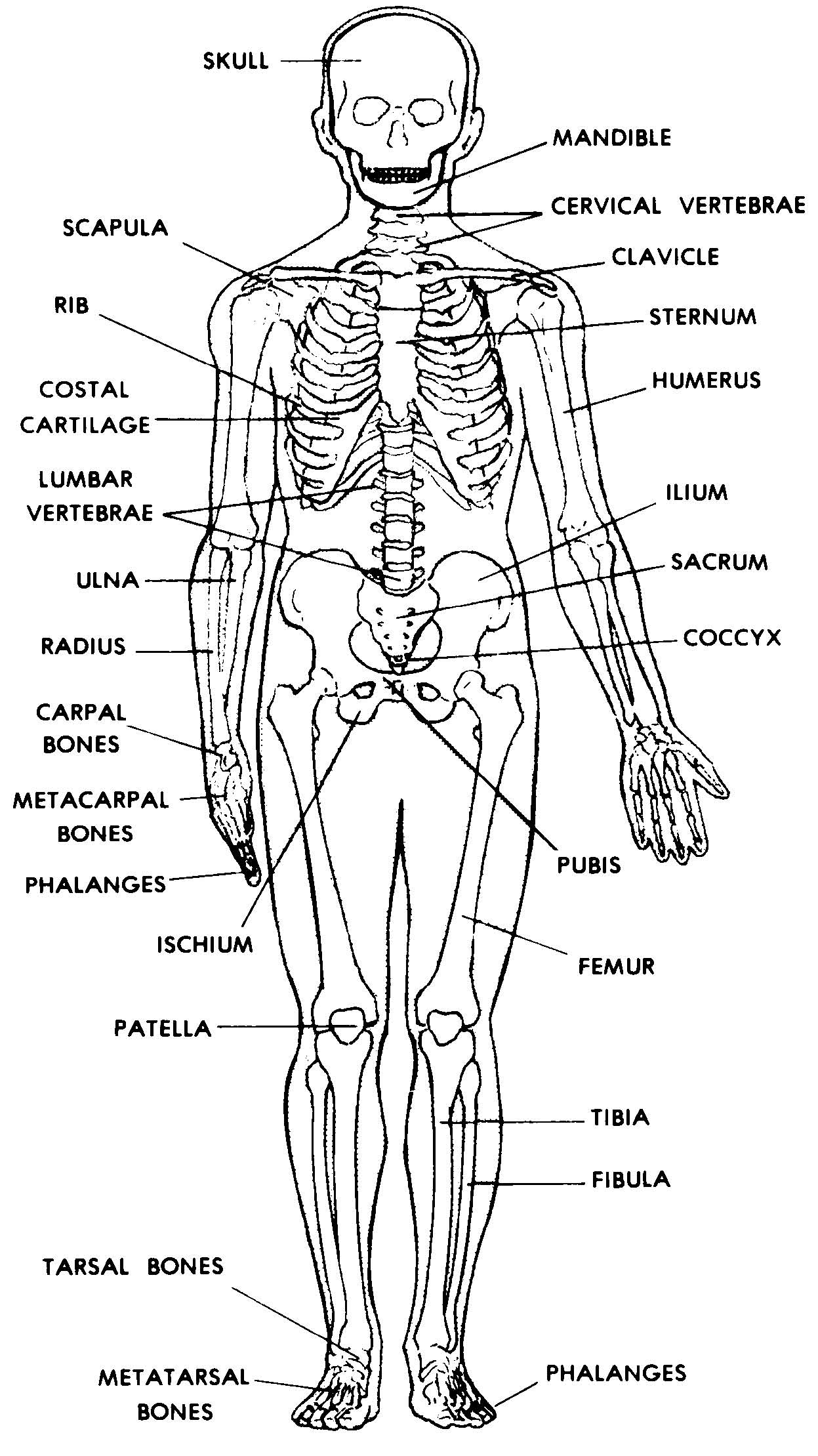
Skeleton Diagram Printable Pictures Human Skeleton Diagram Blank, Human Anatomy Diagram
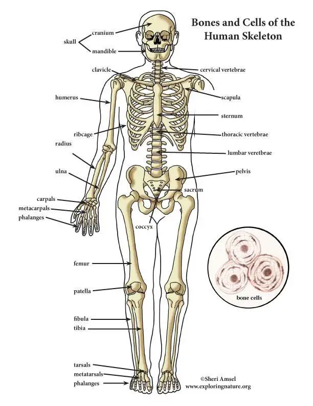
The skeletal system includes all of the bones and joints in the body. Each bone is a complex living organ that is made up of many cells, protein fibers, and minerals. The skeleton acts as a scaffold by providing support and protection for the soft tissues that make up the rest of the body. The skeletal system also provides attachment points for.

Human Skeleton Coloring Page Medical Art Library
Skeletal System. The skeletal system gives the body its basic framework, providing structure, protection, and movement. The 206 bones in the body also produce blood cells, store important minerals.
Human Skeletal System Diagram coordstudenti
Bone is living tissue that makes up the body's skeleton. There are 3 types of bone tissue, including the following: Compact tissue. The harder, outer tissue of bones. Cancellous tissue. The sponge-like tissue inside bones. Subchondral tissue. The smooth tissue at the ends of bones, which is covered with another type of tissue called cartilage.
Skeleton Diagram
skeleton, Bony framework of the body. It includes the skull, vertebral column, collarbone, shoulder blades, rib cage, pelvic girdle and the bone s of the hands, arms, feet, and legs. The skeleton supports the body and protects its internal organs. It is held together by ligaments and moved at the joint s by the muscle s, which are attached to it.
Why do we have bones?
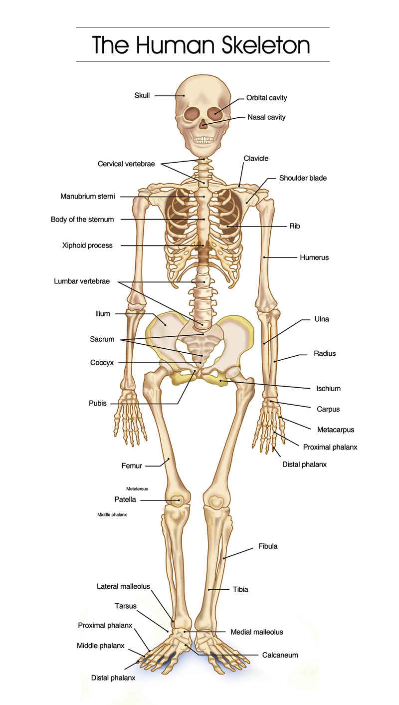
Given below is a labeled diagram, and tips to help you draw and memorize the names of different parts. Human Skeleton Diagram. Here is a detailed diagram which shows the various bones present in an adult skeletal system. There is a little difference between the male and female skeleton, but for diagrams mostly a male skeletal system is considered.